
Especialistas nacionales e internacionales que compartirán su trabajo más reciente en el XIV Congreso Nacional y II Congreso Internacional de Biología del Desarrollo.

Dr. Ariel Bazzini
Stowers Institute for Medical Research
Spatio-temporally regulated maternal RNA decay factor essential for zebrafish development
Maternal mRNA decay is essential for the Maternal-to-Zygotic Transition (MZT), yet its impact on spatial regulation has remained largely unexplored. We identify Cth1 as the first maternally encoded RNA-decay factor regulated in a spatiotemporal manner outside the germline in zebrafish, driven by conserved 3′UTR cis-elements. RfxCas13d-mediated depletion of maternal cth1 revealed early developmental defects that could not be assessed with SpyCas9 mutants due to infertility from impaired gametogenesis. Mechanistically, Cth1 acts independently of zygotic genome activation, recognizing AU-rich motifs in maternal 3′UTRs to promote deadenylation and decay; deletion of these motifs or depletion of Cth1 stabilizes both reporter and endogenous transcripts. These findings establish Cth1 as a key component of the maternal program and provide the first direct evidence that spatially regulated mRNA decay, outside the germline, contributes to early vertebrate development.
Palabras clave
Spatio-temporal maternal RNAs, CRISPR-Cas13d, Zebrafish, MZT
Aspectos destacados
- Cth1 is a highly deposited maternal mRNA that undergoes rapid decay.
- 3’UTR of Cth1 drives its spatio-temporal dynamics.
- Cth1 is essential for gametogenesis and early embryonic development.
- CTH1 recognizes AU-rich elements in target 3′UTRs to trigger decay.

Dr. Roberto Mayor
University College London
Migrar para construir: el viaje colectivo de las crestas neurales
Las crestas neurales son una población celular multipotente cuya migración es esencial para la formación de numerosos tejidos y órganos durante el desarrollo embrionario. En este trabajo exploramos los mecanismos celulares, moleculares y físicos que coordinan su migración colectiva, permitiendo que grupos de células se desplacen de manera eficiente y dirigida. Analizamos cómo las interacciones célula-célula y las señales del microambiente regulan este proceso dinámico. Nuestros resultados aportan nuevas perspectivas sobre los principios que gobiernan los movimientos colectivos celulares y su importancia en la morfogénesis embrionaria.

Dra. Bénédicte Desvoyes
Centro de Biología Molecular Severo Ochoa (CSIC-UAM)
A matter of time: how cell cycle dynamics shape root development
Organogenesis requires a tight coordination between cell proliferation and cell differentiation. Although the genetic networks controlling root development and tissue specification are well described, how they are integrated with cell cycle progression remains unclear. In this talk, I will discuss how cell cycle dynamics are spatially organized in the root meristem, revealing a positional gradient of cell cycle duration. I will also address how developmental regulators contribute to shaping this gradient, linking stem cell maintenance with cell cycle progression. These findings provide a new framework for understanding how growth and patterning are coordinated during root development.

Dr. Fulgencio Alatorre Cobos
Secihiti-Unidad de Biología Integrativa, Centro de Investigación Científica de Yucatán
Análisis de la ontogenia y el desarrollo de hojas compuestas disectadas en angiospermas: el caso de palma jipi, una pseudopalma del Neotrópico
En plantas, el estudio del desarrollo de hojas se ha centrado principalmente en especies con hojas sencillas o lineales, habiendo aún muchas interrogantes sobre cómo se forman aquellas con morfología compleja. En esta charla, compartiremos nuestros avances empleando histología, superresolución de imágenes y transcriptómica espacial para entender el origen y desarrollo de la hoja en palma jipi (Carludovica spp.), una monocotiledónea tropical, con grandes hojas palmeadas disectadas, usadas para elaborar los famosos sombreros de Panamá.

Dr. Edgar Demesa Arévalo
Departamento de Ingeniería Genética, CINVESTAV Unidad Irapuato
Virtual isolation of rare cell types during early meristem establishment in barley
In cereals, the specification of sequential meristematic cell identities and the subsequent establishment of a floral meristem precede flower formation. This complex developmental program requires transcriptional coordination to correctly specify cell lineages. We therefore sought to develop an atlas of the barley meristem featuring single-cell expression data with spatial resolution, with the hope that this atlas will help us to define developmental transitions or cell states with improved resolution.

Dra. María Elena Torres Padilla
Institute of Epigenetics & Stem Cells, Helmholtz Munich
Epigenetic mechanisms in early mammalian development

Dra. M. Fernanda Palominos
UC Berkeley
The ontogenetic basis of craniofacial evolution in pupfishes

Dr. Martín Andrés Estermann
Institute of Molecular Genetics of the Czech Academy of Sciences
Glucose Metabolism as a Driver of Gonadal Sex Differentiation and Fertility
Infertility affects approximately 15% of couples worldwide, with male infertility responsible for nearly half of cases. While dietary and metabolic factors influence reproduction, the molecular mechanisms linking metabolism to fertility remain unclear. Our research explores how cellular metabolism regulates embryonic testis differentiation and its long-term impact on adult fertility using mice. We focus on Sertoli cells, the somatic lineage that supports the germline and orchestrates testis development. Previous studies suggest embryonic Sertoli cells rely on glucose for identity maintenance. Here, we investigated how glucose metabolism regulates Sertoli cell differentiation and function. Using ex-vivo drug screening in embryonic mouse testes, we found that glucose metabolism in Sertoli cells primarily feeds into the hexosamine biosynthetic pathway (HBP) rather than glycolysis. The HBP, through the O-GlcNAc transferase OGT, facilitates protein O-GlcNAcylation, a key post-translational modification that regulates cellular processes, including gene expression and protein stability. To examine its role in testis development, we performed transcriptomic analysis on embryonic testes treated with the OGT inhibitor OSMI-1, revealing defects in Sertoli cell differentiation, testis cord organization, and germ cell survival. To explore this process in-vivo, we generated Sertoli cell-specific Ogt conditional knockout (cKO) mice. Loss of Ogt during the process of sex differentiation destabilized testicular fate and caused ovotestis formation, with reduced SOX9⁺ Sertoli cells and ectopic FOXL2⁺ pre-granulosa cells. Ogt cKO in later stages resulted in infertile mice, with smaller testes, and lacking germ cells in seminiferous tubules, highlighting O-GlcNAcylation’s essential role in Sertoli cell function. Our findings suggest that Sertoli cells prioritize glucose metabolism through the HBP to sustain O-GlcNAcylation, ensuring their differentiation, survival, and germline support. Disrupting this process leads to Sertoli cell dysfunction, germline loss, and ultimately, male infertility.

Dr. Elías H. Barriga
Physics of Life, TU Dresden
The Forces That Shape Us
In this talk, I will discuss how interactions between tissues generate physical scenarios that are sensed, internalized, and translated by molecular machineries to coordinate collective cell behavior. Using collective cell migration in vivo as a framework, I will show how tissue-scale mechanics and bioelectric fields emerge during development, and how cells convert these cues into polarity, migration, and self-organization. Based on the data presented, I hope to stimulate discussion on the idea that robust morphogenesis emerges from feedback between physical tissue states and the molecular programs that fuel, interpret, and translate them into collective cell behaviors.
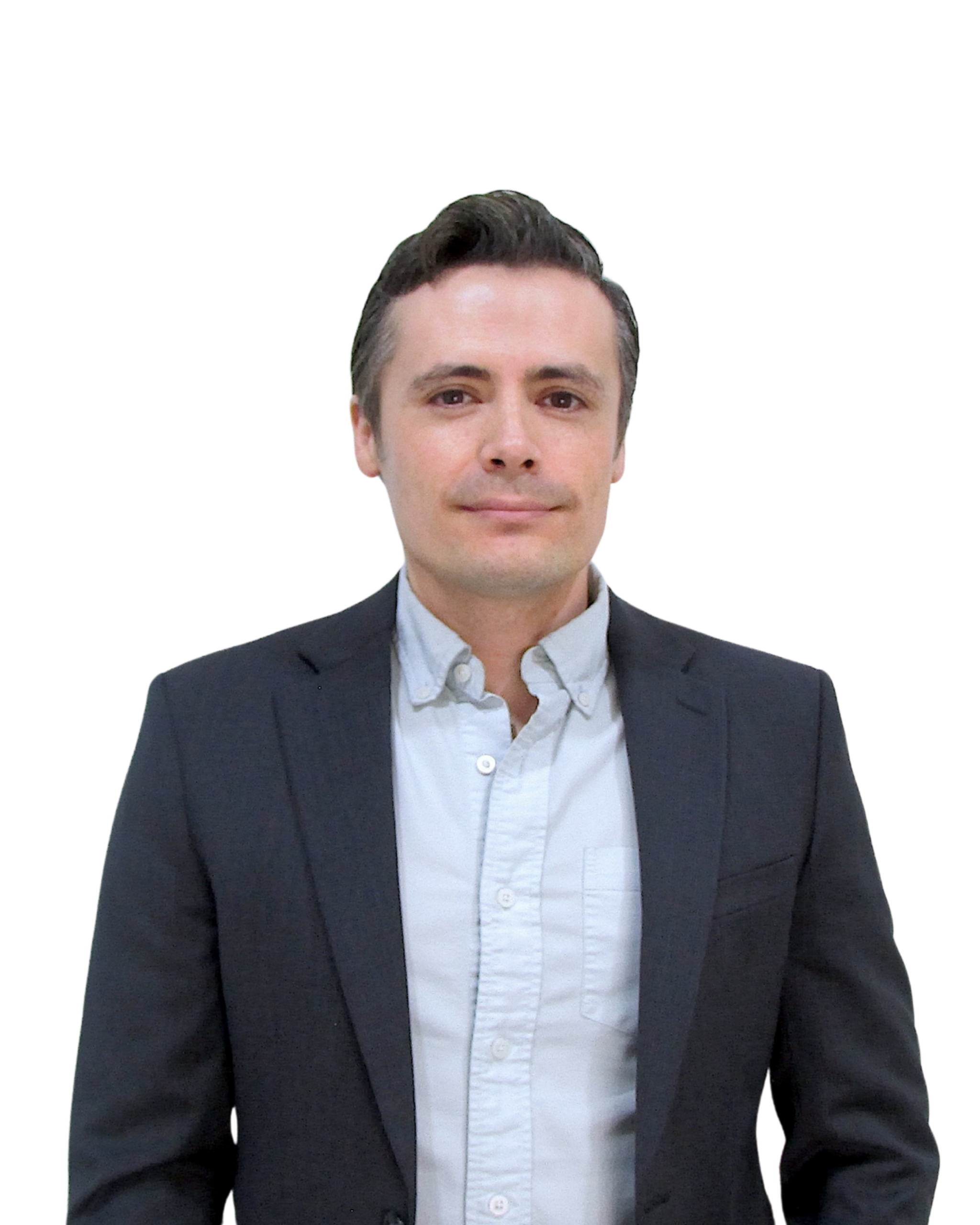
Dr. Sergio E. Campos Rodríguez
Centro de Investigación sobre el Envejecimiento, Sede Sur, CINVESTAV
Biología comparativa del género Hydra para investigar los mecanismos de la longevidad
El aumento de la fragilidad asociado al envejecimiento se atribuye en parte a la disminución de la capacidad regenerativa de los tejidos y órganos. En este sentido, los animales con alta capacidad regenerativa tienden a mostrar un envejecimiento mínimo. El cnidario Hydra vulgaris es capaz de regenerar todo el cuerpo y parece no presentar signos de envejecimiento. Curiosamente, una especie estrechamente relacionada, Hydra oligactis, presenta un fenotipo asociado al envejecimiento en respuesta a la diferenciación sexual. En nuestro laboratorio estamos usando a estas dos especies de Hydra como sistema para abordar el estudio de la longevidad desde una aproximación de biología comparativa. Para ello, estamos caracterizando los signos característicos del envejecimiento en H. oligactis a nivel molecular. Por otro lado, estamos usando estrategias de genómica comparativa para esclarecer la evolución de los genes con un rol en estos signos característicos del envejecimiento a lo largo del árbol de Metazoarios desde una perspectiva del género Hydra, con el fin de entender cómo la longevidad ha sido moldeada a través de la evolución. Más aún, estamos tratando de describir la asociación que existe entre regeneración y envejecimiento usando estas mismas especies de Hydra.

Dra. Marycruz Flores Flores
School of Medicine, University of Colorado
Functional redundancy and divergence of tbx4 and tbx5 genes in zebrafish cardiopharyngeal and lateral plate mesoderm development

Dra. Hilda Lomelí Buyoli
Instituto de Biotecnología, UNAM
El papel del metabolismo de treonina en el desarrollo temprano del pez cebra

Dra. Katarzyna Oktaba
CINVESTAV Unidad Irapuato
Dissecting the regulatory landscape of the dachsous gene during Drosophila development
The atypical cadherin Dachsous (Ds) regulates planar cell polarity (PCP) and organ growth, but its transcriptional control is poorly understood. We identified seven enhancers near the ds promoter that drive expression in ectoderm-derived tissues. While in vivo assays show these enhancers act redundantly during early embryogenesis, CRISPR/Cas9 deletion of a single enhancer produces distinct wing phenotypes in adult flies. These findings offer new insights into the transcriptional regulation underlying Drosophila development.

Dra. Mónica P. Colaiacovo
Harvard Medical School
Germline maintenance and genomic integrity: insights from C. elegans

Dr. Alfredo Varela-Echavarría
Instituto de Neurobiología, UNAM
Studying regeneration in a new experimental model: the garden slug Deroceras laeve
The mantle, foot, body wall, cerebral ganglia, and tentacles are among the many structures that regenerate in terrestrial gastropods. However, the underlying cellular and molecular mechanisms have not been thoroughly studied. Because of its small size, highly complex organs and systems, ease of laboratory culture, and widespread distribution, we developed the garden slug Deroceras laeve as an experimental model for this purpose. We sequenced its genome, assembled it into chromosomes, and annotated repetitive sequences, protein-coding genes and non-coding RNAs. We also built SlugAtlas (slugatlas.lavis.unam.mx), an online anatomical and histological resource for D. laeve and another slug, Ambigolimax valentianus. This allowed a comprehensive comparative analysis of the organs and tissues of these two species and the description of unique and novel features of their anatomy. For D. laeve, we also examined its degrowth and regrowth, which occur in animals with high potential for regeneration, and analyzed the regeneration of its tail and nerve ganglia. Our initial findings reveal morphological features, cellular mechanisms, and gene pathways that have been previously linked to comparable processes in diverse animal groups, as well as others that have not been connected to tissue restoration. These achievements lay the groundwork for using the slug D. laeve to understand the complex mechanisms behind gastropod regeneration.
Financiamiento
DGAPA-PAPIIT-UNAM IN204526, SECIHTI CBF2023-2024-834

Dr. Juan Tena
Centro Andaluz de Biología del Desarrollo (CABD)
Hematopoyesis primitiva en el cerebro adulto del killifish
Aunque el envejecimiento es un proceso sistémico que afecta a todo el organismo, el cerebro constituye uno de los órganos más vulnerables y está sujeto a numerosas enfermedades neurodegenerativas. En este estudio, analizamos los tipos celulares y los genes particularmente susceptibles al proceso de envejecimiento. Para ello, realizamos una serie de experimentos de RNA-seq de célula única a lo largo del tiempo en el cerebro del killifish, un modelo vertebrado con una esperanza de vida muy corta, lo que lo posiciona como un modelo idóneo para el estudio del envejecimiento. Nuestro análisis revela que las células progenitoras no gliales se encuentran entre las poblaciones que experimentan cambios más significativos entre los individuos jóvenes y los de edad avanzada. Además, identificamos clústeres estromales especializados que parecen sustentar un programa hematopoyético primitivo en el cerebro, el cual, en otras especies vertebradas, se encuentra activo únicamente durante las etapas embrionarias. Los resultados obtenidos sugieren que el mantenimiento de programas genéticos embrionarios es una característica general del cerebro adulto del killifish. Es probable que dichos programas contribuyan a sostener las elevadas tasas de proliferación y metabolismo propias de esta especie, así como a contrarrestar el daño celular acumulado durante las etapas avanzadas de la vida.

Dr. Acaimo González-Reyes
Centro Andaluz de Biología del Desarrollo (CABD)
RNA and stem cells come together: on splicing and translational repression
Stem cell exhaustion is a hallmark of ageing, yet how age-associated changes in stem cell niches impair their ability to sustain tissue regeneration is not fully understood. The Drosophila ovarian niche provides a powerful model to investigate how ageing reshapes the molecular landscape of a stem cell niche. We have shown that niche ageing is accompanied by extensive, cell type-specific changes in gene expression and alternative splicing, highlighting post-transcriptional regulation as a key component of niche decline. Different populations of niche support cells undergo distinct molecular ageing trajectories, suggesting that niche dysfunction arises through cell type-specific mechanisms. In addition, we identified the splicing regulator Smu1 as essential for niche function and germline stem cell maintenance, acting through alternative splicing and localization of the cell-adhesion molecule Fasciclin 2 (Fas2). I will also present the unexpected role of the translational repressor Bruno 1 (Bru1) as a key regulator of the mitosis-to-meiosis transition in female gametogenesis. Loss of Bru1 causes accelerated mitoses, DNA damage, cellular stress and apoptosis. Together, our findings establish post-transcriptional regulation as an important driver of tissue ageing and germline homeostasis, linking age-associated changes in RNA processing and translation to altered cell function, niche integrity and stem cell maintenance.

Dra. Diana Escalante-Alcalde
Instituto de Fisiología Celular, UNAM
Relevancia de la Regulación de Lípidos Bioactivos en el Desarrollo de la Cresta Neural
Las células de la cresta neural (CCN) son una población multipotente que genera una gran variedad de tipos celulares y estructuras en vertebrados. Por lo anterior, alteraciones en este linaje pueden originar un amplio grupo de enfermedades en el humano conocidas como neurocristopatías. La señalización de factores proteicos durante el desarrollo de las CCN se ha estudiado ampliamente, sin embargo, se ha dado poca atención a la señalización mediada por lípidos bioactivos. La fosfatasa de fosfolípidos-3 (PLPP3) es una proteína con la capacidad de regular la concentración de al menos 4 fosfolípidos bioactivos y por lo tanto sus actividades biológicas. La inactivación condicional de Plpp3 en las CCN del ratón produce letalidad neonatal acompañada de dismorfología craneofacial y alteraciones en la función cardiaca. Los neonatos mutantes presentan alteraciones en el ritmo cardiaco y defectos en la cobertura de los nervios simpáticos que inervan al corazón. Además, experimentos de scRNA-seq mostraron alteraciones en la transcripción de genes asociados a la persistencia del ducto arterioso (PDA), fenotipo que fue posteriormente confirmado. Aun desconocemos si la letalidad se debe sólo a la PDA o al efecto aditivo de la falta de innervación simpática. Las alteraciones craneofaciales están representadas por desplazamiento anterior de la posición de la sutura coronal del cráneo e hipoplasia de la región frontonasal acompañada de hipotelorismo. Estos defectos están asociados a un incremento en la muerte de las CCN que participan en la formación de dichas estructuras o a alteraciones en las interacciones con las células del mesodermo con el que colindan. Nuestros resultados resaltan la importancia que tiene la función de esta enzima en la biología de las CCN y abre un nuevo campo para estudiar defectos en el desarrollo de la cresta neural en el humano.
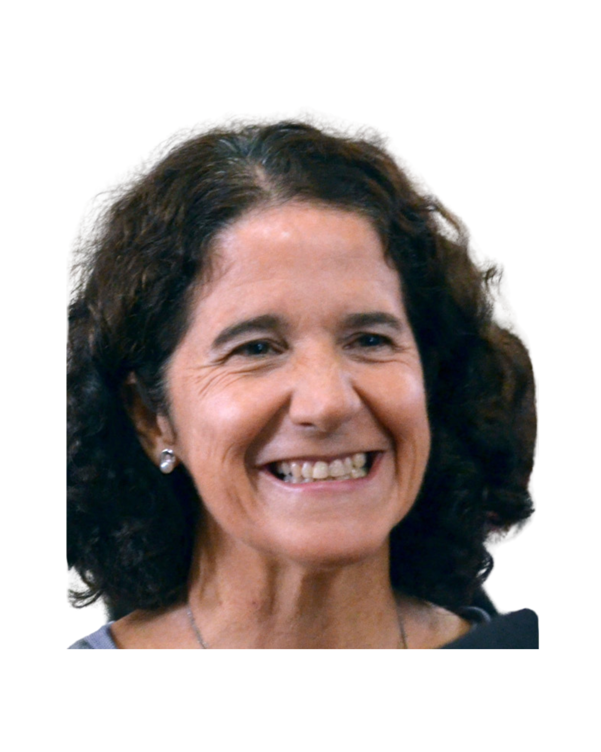
Dra. María Dolores Martín Bermudo
EMBO Keynote Lecture
When Cells Feel the Matrix: Shaping Tissues and Maintaining Homeostasis
Más ponentes confirmados
- Dr. Isaac Skromne University of Richmond, US